Pelvis Muscles Mri Anatomy - Soleus Muscle Radiology Reference Article Radiopaedia Org : The muscle originates from the body of the pubis and attaches to the pectineal line and proximal part of the linea aspera of femur.
byAdmin•
0
Pelvis Muscles Mri Anatomy - Soleus Muscle Radiology Reference Article Radiopaedia Org : The muscle originates from the body of the pubis and attaches to the pectineal line and proximal part of the linea aspera of femur.. The muscles of the pelvic floor are collectively referred to as the levator ani and coccygeus muscles. Magnetic resonance imaging (mri) utilizes magnet and radio waves to produce diagnostic images that allow a doctor to visualize the hips. Functional anatomy of the male pelvic floor online course: Thigh muscles are responsible for allowing normal gait and proper lower extremity function (1). Anatomy of the female pelvis mri atlas of the human body using cross sectional imaging.
This mri male pelvis axial cross sectional anatomy tool is absolutely free to use. The pelvic diaphragm is composed of the ischiococcygeus muscle and levator ani muscle, the latter of which consists of the iliococcygeus, puborectalis, and pubococcygeus muscles. The medial thigh muscles are responsible for the adduction (movement of a body part toward the body's midline) of the leg. This anatomy section promotes the use of the terminologia anatomica, the international standard of anatomical nomenclature. They form a large sheet of skeletal muscle that is thicker in some areas than in others.
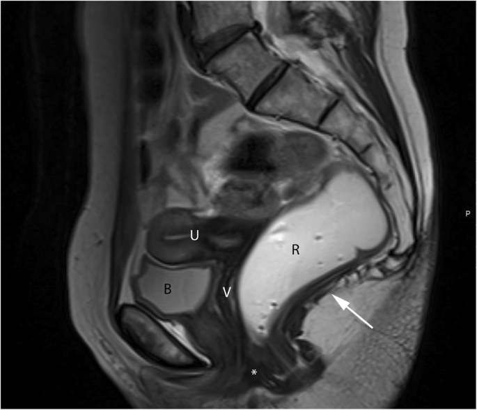
Dynamic Magnetic Resonance Imaging Of The Female Pelvic Floor A Pictorial Review Insights Into Imaging Full Text from media.springernature.com Pelvic mri anatomy553 lower uterine segment; Magnetic resonance imaging (mri) devices can provide direct transverse, sagittal, and coronal plane images. The piriformis is a flat muscle, pyramidal in shape, lying almost parallel with the posterior margin of the gluteus medius muscle and deep to the gluteus maximus muscle. This anatomy section promotes the use of the terminologia anatomica, the international standard of anatomical nomenclature. Use the mouse scroll wheel to move the images up and down alternatively use the tiny arrows (>>) on both side of the image to move the images.>>) on both side of the image to move the images. Duplicated cervices are present.18on mri, both entities are characterized by a split uterine horns that demonstrate zonal uterine anatomy. The pubococcygeus muscle runs from the inner surface of the pubis and obturator fascia with fibers fusing medially at the perineal body and musculature of the prostate / vagina. 47 adductor magnus muscle this is the largest of the group of adductors of the thigh.
47 adductor magnus muscle this is the largest of the group of adductors of the thigh.
The location of the urogenital diaphragm is caudal to the pelvic diaphragm and anterior to the anorectum. They form a large sheet of skeletal muscle that is thicker in some areas than in others. It is situated partly within the pelvis against its posterior wall, and partly at the back of the hip joint. The innominate bones articulate with each other anteriorly and with the sacrum posteriorly. It provides an interpretative algorithm for approaching an unknown pelvic lesion at mri. Mri anatomy of the male pelvis and pelvic floor in multiple patients aged 50 years or older. See pelvic fractures (summary) and pelvic fractures The pelvic cavity and perineum. This mri hip joint axial cross sectional anatomy tool is absolutely free to use. The thigh has some of the body's largest muscles. Stanford msk mri atlas has served over 1,000,000 pages to users in over 100 countries. Knee shoulder shoulder arthrogram ankle elbow wrist hip contact. Please email baodo at stanford.edu.
This imaging technique is used to diagnose and evaluate edema, partial or complete muscle tears, and hematoma. The labeled structures are (excluding the correct side): Anteriorly, pubocervical fibromuscularis is attached to the levator ani using arcus tendineus fascia pelvis (fig. Pelvic mri anatomy553 lower uterine segment; 47 adductor magnus muscle this is the largest of the group of adductors of the thigh.
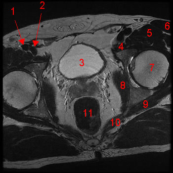
Male Pelvis Magnetic Resonance Imaging W Radiology from w-radiology.com The muscle originates from the body of the pubis and attaches to the pectineal line and proximal part of the linea aspera of femur. This mri male pelvis axial cross sectional anatomy tool is absolutely free to use. Dotted line in a ) show the anatomy of the. Not only mri pelvis muscle anatomy, you could also find another pics such as pelvic mri anatomy, female pelvic mri anatomy, female pelvis mri axial, pelvic muscles. Abdominal and pelvic anatomy encompasses the anatomy of all structures of the abdominal and pelvic cavities. The muscles of the pelvic floor are collectively referred to as the levator ani and coccygeus muscles. The different views are advantageous to clinicians assessing different pathologies related to the male pelvis(7). Duplicated cervices are present.18on mri, both entities are characterized by a split uterine horns that demonstrate zonal uterine anatomy.
Magnetic resonance imaging (mri) devices can provide direct transverse, sagittal, and coronal plane images.
Anatomy of the female pelvis mri atlas of the human body using cross sectional imaging. It is situated partly within the pelvis against its posterior wall, and partly at the back of the hip joint. This article highlights the normal anatomy of the pelvic spaces in the female pelvis and focuses on mri features of common tumors and tumor mimics that arise in these spaces. This imaging technique is used to diagnose and evaluate edema, partial or complete muscle tears, and hematoma. Mri anatomy of the male pelvis and pelvic floor in multiple patients aged 50 years or older. Magnetic resonance imaging or mri of the female pelvis offers a unique display of the pelvic anatomy, including a woman's ovaries, uterus, and fallopian tubes. Magnetic resonance imaging (mri) devices can provide direct transverse, sagittal, and coronal plane images. Mri provides superior soft tissue contrast resolution for imaging the anatomy best seen in t1 weighted and pathology best seen on t2 weighted of the pelvis 3. The piriformis is a flat muscle, pyramidal in shape, lying almost parallel with the posterior margin of the gluteus medius muscle and deep to the gluteus maximus muscle. This mri hip joint axial cross sectional anatomy tool is absolutely free to use. The pelvis consists of an osseous ring formed by the innominate bones and sacrum, with numerous muscles for support. Functional anatomy of the male pelvic floor online course: The key feature is a deep concavity of the expected uterine fundus, with a cleft of at least 1 cm diagnostic.
Invasive angiography is the gold standard modality for assessing pelvic vasculature 3. See pelvic fractures (summary) and pelvic fractures Pertaining to mri of the pelvic floor is oriented toward evaluation of the female pelvic floor. Knee shoulder shoulder arthrogram ankle elbow wrist hip contact. Dotted line in a ) show the anatomy of the.
Normal Shoulder Mri How To Read A Shoulder Mri Kenhub from thumbor.kenhub.com The iliococcygeus muscle attaches to the inner tip of the coccyx posteriorly. The levator ani has three main components, each of which is paired 1,2,5: Anatomically, the pelvis can be divided into true and false pelvis by an oblique line that extends from the sacral promontory along the anterior aspect of s1 to the symphysis pubis. Duplicated cervices are present.18on mri, both entities are characterized by a split uterine horns that demonstrate zonal uterine anatomy. Use the mouse scroll wheel to move the images up and down alternatively use the tiny arrows (>>) on both side of the image to move the images.>>) on both side of the image to move the images. Mri anatomy of the male pelvis and pelvic floor in multiple patients aged 50 years or older. Abdominal and pelvic anatomy encompasses the anatomy of all structures of the abdominal and pelvic cavities. It is situated partly within the pelvis against its posterior wall, and partly at the back of the hip joint.
Anatomy of the abdomen and male pelvis using cross sectional imaging ct interactive atlas of human anatomy we have created an anatomical atlas of abdominal and pelvic ct which is an interactive tool for.
The muscle originates from the body of the pubis and attaches to the pectineal line and proximal part of the linea aspera of femur. Mri anatomy of the male pelvis and pelvic floor in multiple patients aged 50 years or older. Pelvic mri anatomy553 lower uterine segment; Anatomically, the pelvis can be divided into true and false pelvis by an oblique line that extends from the sacral promontory along the anterior aspect of s1 to the symphysis pubis. Functional anatomy of the male pelvic floor online course: Anatomy of the abdomen and male pelvis using cross sectional imaging ct interactive atlas of human anatomy we have created an anatomical atlas of abdominal and pelvic ct which is an interactive tool for. The levator ani has three main components, each of which is paired 1,2,5: Not only mri pelvis muscle anatomy, you could also find another pics such as pelvic mri anatomy, female pelvic mri anatomy, female pelvis mri axial, pelvic muscles. Use the mouse scroll wheel to move the images up and down alternatively use the tiny arrows (>>) on both side of the image to move the images.>>) on both side of the image to move the images. The muscles of the pelvic floor are collectively referred to as the levator ani and coccygeus muscles. Mri pelvis anatomy free male pelvis axial anatomy 15 liver 16 oesophagus 17 stomach. This anatomy section promotes the use of the terminologia anatomica, the international standard of anatomical nomenclature. Dotted line in a ) show the anatomy of the.
The pubococcygeus muscle runs from the inner surface of the pubis and obturator fascia with fibers fusing medially at the perineal body and musculature of the prostate / vagina anatomy muscles pelvis. Please email baodo at stanford.edu.